INTRODUCTION
Since the Zollner [1] and Wullstein [2] first reported their experiments about reconstruction of the hearing, various graft materials and methods have been used to close tympanic membrane perforation such as fascia, vein, dura or cartilage in overlay or underlay fashion [3]. Either endoscopic or open microscopic approach can be used in each technique, although usually it is still being performed via microscopes rather than endoscopes despite canal incision or ear packing. In simple chronic otitis media (COM) sequelae more limited incisions and tympanic membrane closure techniques had also been investigated widely. Eventually, Eavey [4] introduced cartilage butterfly inlay tympanoplasty technique in children for closure of small to medium sized central perforations in 1998. Because this technique does not require tympanomeatal flap elevation and it can be performed under local anesthesia, less operating time, minimal scarring due to transcanal approach, lack of ear packing, simple follow-up period can be obtained when compared with other tympanoplasty techniques. There are three difficulties related to classic butterfly cartilage tympanoplasty technique: (1) anterior manipulation and fitting difficulty of the cartilage because of insufficient vision; (2) graft stabilization challenges, particularly the eustachian tube function; and (3) exact measurement of the perforation size. These factors are strongly associated with surgical and hearing outcomes in butterfly technique. Furthermore, age, surgeon experiment, and perforation size may also influence hearing outcomes [5].
Butterfly cartilage tympanoplasty was evaluated in the terms of (1) presence of the graft resorption; (2) residual perforation; (3) iatrogenic cholesteatoma and epithelial pearl; and (4) long-term outcomes of the hearing. Although these instances are concerns about butterfly tympanoplasty, there is no such study investigating all of them. Most of the studies in the literature report short-term results whereas long-term results are necessary to evaluate the efficiancy of the butterfly tympanoplasty. Our study has longer follow-up periods and larger patient population compared with the other studies in the literature. The purposes of this study were to evaluate and report the long-term results of the butterfly cartilage tympanoplasty and investigate the concerns as mentioned above. Short-term and long-term hearing outcomes were compared according to age and perforation location as well.
MATERIALS AND METHODS
We retrospectively evaluated the medical records of patients who underwent butterfly cartilage tympanoplasty at an otolaryngology department of a tertiary medical center between February 2006 and December 2014. Microscopic evaluation was performed in all patients at 1st week, 1st month and 6th month and 24th month after surgery. Additionally, pure tone audiometry (PTA) was measured at 6th and 24th month follow-up. Age, gender, follow-up time, pre- and postoperative PTA thresholds (both air and bone conduction), pre- and postoperative air-bone gaps (ABGs), if any residual perforation were noted. All procedures performed in studies involving human participants were in accordance with the ethical standards of the Ege University Institutional Review Board (IRB No. 54698) and/or national research committee and with the 1964 Helsinki declaration and its later amendments or comparable ethical standards. Informed consent was obtained from all individual participants included in the study.
Patients
A total of 93 of 108 patients (50 males and 43 females) aged 15 to 75 years were enrolled in this study who had at least 2 years follow up and those that (mean follow-up period, 41.8 months). All data were obtained from patientŌĆÖs folders and hospital information system. Ninety-three patients were included in this study who had exact data such as PTA, follow-up findings, endoscopic records. Fifteen patients were excluded due to lack of follow-up. Patients those followed regularly and PTA was measured at 6th and 24th month follow-up were included in this study. Patients who had not regularly follow-up period were excluded. Study involved patients who underwent butterfly cartilage tympanoplasty due to anterior, posterior, and central tympanic membrane perforation, except marginal perforation. The mean age was 40.3 years. Surgery was performed under general anesthesia in 9 patients and local anesthesia in 84 patients. Inclusion criteria for this study were (1) patients who had regular follow-up and (2) PTA measurement at 6th and 24th month follow-up. Perforations were divided into three categories; anterior, posterior, and central. Moreover, the size of perforation was observed and classified into two groups. Tympanic membrane (TM) perforation which was between 20% and 40% as small and between 40% and 60% of the TM as medium size perforation.
Patients with perforations greater than 60% to total of the TM, extensive myringitis, history of otorrhoea within the last month, possible mastoid cell pathology requiring exploration of the middle ear such as cholesteatoma, discordant hearing loss with the size of perforation, atrophied tympanic membrane or retraction pocket, marginal perforations, possible ossicular chain problem did not include the study. Conversely, patients with dry ear at the time of surgery, absence of cholesteatoma, absence of tympanic membrane atelectasis underwent butterfly tympanoplasty.
Pure tone audiometry
PTA was measured prior to surgery and at postoperative 1st, 6th, 12th, 24th month follow-up. Standard PTA (Interacoustic AC-40, Interacoustics, Middelfart, Denmark; headphone: TDH39) was performed before and after the surgery for the frequencies of 0.5, 1, 2, 4 kHz. Pre- and postoperative PTA thresholds at 0.5, 1, 2, 4 kHz were noted. The ABG was calculated in both pre- and postoperative PTA.
Surgical technique
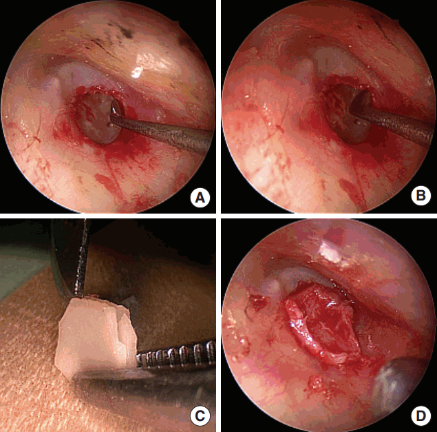
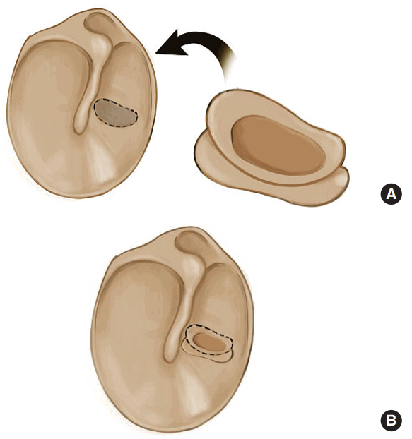
All patients were operated by the same senior surgeon (CB). Surgery was performed under general anesthesia or local anesthesia. Transmeatal approach was used in all patients with the use of microscope. Following patient preparation, lidocaine and epinephrine 1:100,000 were used for the infiltration of the canal skin and meatal surface of the tragus. The lateral 2-mm portion of the tragal cartilage was left intact in the dome of the tragus for cosmesis. Tragal cartilage was harvested without perichondrium on either surfaces. The incision was closed with 5/0 polypropylene sutures. Subsequently circumferential marginal trimming to freshen up the tympanic membrane was made with a straight pick. Sufficient TM remnant was left in place to secure graft within the TM. Dimension and shape of the perforation were measured with plester micro ear knife (Sklar Surgical Instruments, West Chester, PA, USA). The same plester was used to shape harvested cartilage according to perforation. A horizontal groove along the border of the graft was made circumferentially at a depth of 1 to 2 mm via 11 blade scalpel in order to imitate the wings of a butterfly. The graft is nestled into the edge of the perforation margin so that one butterfly wing remains lateral and the other wing is on the medial side of the perforation (Fig. 1). The graft was placed with a pick similar to a grommet tube insertion. Patients were examined routinely at 1st week, subsequent visits were carried out at 3, 6, 12, and 24 months after surgery. At each follow up, patients were examined under microscope. Any complications were noted such as otitis externa, otitis media, graft displacement, and retraction of the TM. PTA was also measured at 2 years follow-up (Fig. 2).
Statistical analysis
Statistical analysis was made using IBM SPSS ver. 22.0 (IBM Corp., Armonk, NY, USA). Chi-square exact test was used for the comparison of categorical data while independent and paired samples t-test were used for the analysis of parametric variables, Wilcoxon and Mann-Whitney U-tests were used for the analysis of nonparametric variables based on the distribution pattern of the data. The distribution pattern was determined by Shapiro-Wilk test. Correlation analysis was performed via Spearman or Pearson correlation analysis depending on the type of the variable. Data were expressed as mean┬▒standard deviation (SD), percentage, range, 95% confidence interval (CI), and median (interquartile range [IQR]) where appropriate. The P<0.05 was considered as statistically significant.
RESULTS
A total of 93 patients underwent transcanal butterfly cartilage tympanoplasty. The mean age was 40.3 years and the mean follow-up period was 41.8 months (range, 24 to 105 months). At the end of the follow-up period, successful closure occurred 88 in 93 patients and success rate is 94.6%. We failed in 5 patients, 3 of them had residual perforation and 2 had total graft resorption. Of the 5 patients, 3 had anterior and 2 had posterior residual perforation. Fat graft myringoplasty was performed in three patients, who had anterior and partial graft failure within 6 months after surgery under local anesthesia and open tympanoplasty was performed 1 year after surgery in the remaining 2 patients because of the lack of residual membrane on the one side of the perforation. All patients no longer had residual perforation after fat graft myringoplasty. All patientsŌĆÖ demographics and distributions among groups are listed in Table 1. There was no significantly difference between groups according to distribution of the age, gender, follow-up period, and perforation location (Table 1).
In all patients, included graft failures, the mean preoperative bone conduction threshold was 15.9 dB (range, 5 to 50 dB) among all groups whereas mean air conduction threshold was 36.4┬▒15.1 dB (range, 10 to 90 dB) preoperatively and 28.8┬▒ 14.3 dB in 6th month follow-up and 24.9┬▒14.1 dB 24th month follow-up (Fig. 3). Preoperative mean ABG was 22.1┬▒7.1 dB (range, 5 to 40 dB) whereas 13.3┬▒5.9 dB 6 months after surgery and 11.9┬▒5.5 dB 24 months after surgery. Pre- and postoperative hearing results and comparison between groups (Table 2). There was significantly difference between pre- and postoperative ABG in both 6th and 24th month follow-up (P6 mo-24 mo<0.05). Furthermore preoperative mean air conduction differed significantly from postoperative 6 improvement was 5.3 in small perforations and 8.6 in medium sized perforations. There was significantly difference between two groups according to ABG (P=0.034). According to perforation location, distribution of the hearing alteration shows in Table 3. Mean ABG did not differ significantly within the each group (Psmall perforation=0.729 and Pmedium perforation=0.481) however, according to perforation location ABG improvement differed significantly between small and medium perforations (Panterior<0.05, Pposterior=0.03, and Pcentral<0.05). Summary of the hearing outcomes are shown in Table 4.
ABG improvement was not significantly different when compared with the age. Patients were divided into two groups, smaller than 40 years age and greater than or equal to 40 years age. Short- and long-term ABG improvement was similar to each other between ages (Pshort-term =0.112 and Plong-term =0.957) whereas mean ABG showed strongly difference between ages (P<0.05).
In patients who had residual perforation and underwent fat graft myringoplasty and open tympanoplasty short-term ABG improvement was 13.2 (whereas 7.16 in whole medium sized perforations-obtained from PTA which was measured prior to 2nd surgery) and long-term ABG improvement was 16.4 (12.89 in total of medium perforations) dB (PTA was measured 2 years after first surgery). Mean pre- and postoperative ABG was 23 and 6.6 respectively (P<0.05).
No case of any complications was recorded like hearing loss, otitis externa, otitis media or graft displacement, retraction of tympanic membrane. Nevertheless, we saw myringitis after the surgery in 4 patients, but no medical treatment was required in these patients. We observed that the complaints spontaneously declined in these patients.
DISCUSSION
Treatment of simple COM varies and it includes open and endoscopic approaches. These techniques require incision or tympanomeatal flap elevation thus postoperative pain, wound healing and ear packing are the disadvantages of these techniques. However, butterfly cartilage tympanoplasty is lack of incision and flap elevation so reduced postoperative pain, shorter healing time can be obtained. Butterfly cartilage tympanoplasty is a relatively new technique based on clinical and experimental findings of the use of cartilage grafting [1,5,6]. The main advantages of the butterfly cartilage technique are its reliability, absence of tympanomeatal flap elevation that may extend surgical time and may be challenging with myringosclerotic ear drums and anterior perforations, shorter operative time, and minimal postoperative discomfort. Although the vast majority of previously published studies which involve hearing outcomes, modified techniques and success rates, report similar results, somehow only a few of them investigate all together.
Butterfly cartilage tympanoplasty is a technique with high success rates. Several other studies have also reported similar rates [1,7-10]. Bhattacharya et al. [9] reported 93.3% success rate, whereas Mauri et al. [10] reported 88.2% success rate. We found a similar success rate for small and medium sized perforations in our study (94.6%). We did not observe graft failure in any of our cases with small perforations, whereas there was minimal residual perforation in 2 cases and graft was totally resorbed in 3 patients with medium sized perforations. Three patients underwent open tympanoplasty due to lack of the tympanic membrane remnant. Among all cases that developed residual perforation, perforation was greater than 50% of the TM at the time of the first surgery. Two of these cases underwent fat graft myringoplasty at the 6th month, while others underwent open tympanoplasty at the end of the 1st year. Interestingly, when these patientsŌĆÖ ABG compared with pre- and postoperative 2nd year, improvement of ABG in patients who need revision surgery, the improvement in their ABG was found to be greater than patients who did not need revision. This may be because all these patientsŌĆÖ perforations were greater than the other perforations, resulting in slightly more ABG, and therefore, greater gain. Additionally, this may be due to very few numbers of patients in this group.
Although theoretically, it is possible for a superficial layer of the TM to migrate to the middle ear via graft wings, we did not observe iatrogenic cholesteatoma in the present study. Also, previous studies did not report any cholesteatoma related with butterfly tympanoplasty [4,10,11]. It is a reliable method in this regard. Nevertheless, careful replenishment of the perforation edges will eliminate the risk of iatrogenic cholesteatoma and long-term follow-up is necessary to observe any clue about this. In this regard our follow-up time is the longest duration in the literature therefore reliability of the findings are superior to most of the previous studies and it was considered adequate to expose long-term complications such as cholesteatoma [1,3,12].
Alain et al. [7] examined hearing gains following total, subtotal and annular perforations, and they reported an average gain of 27.1 dB. Riss et al. [13] reported a gain of 5.8 dB, and Ulku [14] reported a gain of 11.28 dB. In our study, preoperative PTA average improved from 36.4 dB to 28.8 dB. Although this result was similar to the findings of Riss et al. [13] and Ulku [14], it is much behind the gain reported by Alain et al. [7] which was 27.1 dB. This may be because Alain et al. [7] included total-subtotal perforations in their study, and these patients have greater hearing loss compared to small-medium perforations. Additionally, average gains in our patients were further increased according to PTA at the 2nd year. The average in 2nd year PTA regressed down to 24.9 dB. This means approximately a gain of 12 dB. Moreover, based on ABG, we observed a gain of 9 dB at the 6th month and a gain of 11 dB at the 24th month. The reason why there was the difference between the 6th month and 2nd year gains may be that graft thickness was reduced and it accommodated to the TM, enhancing its elasticity. Obviously, more studies are needed to clarify this. Additionally, with regard to the perforation size, while average ABG improvement was 5.3 dB in small perforations, it was 8.6 dB in moderate perforations. However, regarding ABG improvements obtained at the 24th month, this gain is observed to increase up to 12.9 dB in moderate perforations. This explains the high gain reported by Alain et al. [7]. For this reason, in order to assess the success of this technique, PTA performed at the late rather than early period seems to be of greater value. During counseling with patients, this progressive increase in the gain should be mentioned, and both the surgeon and the patient should show patience for evaluating the outcomes. In this respect our study is the first trial which has the largest patient group in the literature that compares 6 and 24 months follow-up results.
Other than the perforation size, its localization can also determine the obtained hearing gain and hearing loss. However, Mehta et al. [15] reported that localization of perforation did not have any influence on hearing loss. Also, Park et al. [16] did not find an association between perforation localization and ABG. In the present study, we also did not find any association between perforation localization and ABG and hearing gains. Although butterfly cartilage tympanoplasty is available in small and medium sized perforations, open tympanoplasty can be used in large anterior perforations because of the restricted anterior vision. Furthermore because of the lack of anterior remnant, graft placement may be somewhat difficult and inadequate thus intact eustachian tube function may cause graft displacement in large anterior perforations.
Apart from these, Tos [17] reported that highest success rates with type 1 cartilage tympanoplasty were observed among patients at or below 10 years old. In contrast, Kessler et al. [18] and Rozendorn et al. [19] reported that graft failure was more frequent among patients younger than 6 and 9 years old. In the present study, mean patient age was 40.3 years age, ranging between 12 and 75 years. Patients who developed graft failure were aged between 17 and 66 years. Regarding hearing gains, we did not find any difference between patients who were smaller than 40 years age and greater than or equal to 40 years age. This indicates that age has no effect on graft failure or hearing gain. In addition to these, apart from the development of residual perforation in 5 patients, we did not observe postoperative complications (otitis externa, otitis media, cholesteatoma, retraction, etc.) in our patients.
Disadvantages of this procedure: (1) Technique is a relatively difficult for beginners, butterfly myringoplasty requires precise evaluation of the perforation size and shape, meticulous preparation of the edges, and exact sizing of the graft; (2) It can be difficult to fit a large graft in a small perforation, even though a smaller graft can be unstable with larger perforation and result in an unsuccessful outcome; (3) Large size of perforations cannot be treated with this technique; (4) Furthermore, if there is no enough membrane remnants in the edges of perforation such as annular perforations it is difficult to success the procedure; (5) We canŌĆÖt sufficiently evaluate middle ear structures and ossicular chain pathologies in this procedure; and (6) Theoretical disadvantage of cartilage is that it makes an opaque tympanic membrane posteriorly, which could potentially hide disease [8].
The retrospective study design was the major limitation of the present study. Nonetheless, the patient number was greater than many of the previous studies. For that matter, the hearing gain could be compared in relation to perforation size and localization with greater credibility. In addition, there are few studies that perform a comparison of all the results.
In conclusion, there are many surgical treatment options for COM. Incision-free or minimally invasive techniques have been preferred at an increasing rate within the recent years. As one of these methods, butterfly tympanoplasty is effectively used in treating simple COM. Although it is challenging to perform in some situations, it can be safely performed in small, moderate, and even large perforations, as the hearing outcomes and successful closure rate are similar to those of other surgical methods. Moreover, it can be performed under local anaesthesia and it has low complication rates. Hearing gains are greater with greater perforation size, but it has no association with perforation localization. Additionally, late period PTA should be preferred instead of early period PTA for assessment of hearing gains.