INTRODUCTION
Thyroid surgery is one of the most common operations among head and neck surgeries. Although this surgery has been performed for many years, recurrent laryngeal nerve (rln) paralysis is still one of the most critical complications and its rate following thyroidectomy is about 1% to 2% [1]. The damage on the nerve could reach up to 14% [2,3].
Knowing the full anatomy of this nerve is the most critical issue in surgery. The nerve on the right side follows a path down the anterior surface, then up the posterior surface of the subclavian artery and reaches the trachea at the level of thyroid gland lower pole [4-7]. The left side of the nerve crosses the arcus aorta and turns up to reach trachea at the level of thyroid gland lower pole [4-7]. While the right rln follows a more oblique way, the left rln is more closer to trachea [4-7]. Until the point where they cross the inferior thyroid artery, right and left rln rise mainly through tracheoesophageal groove [5-7]. They may cross the artery in the front, back or in between its branches [4,8,9]. On both sides before reaching the Berry ligament, rln forms bifurcation [5,6,8]. Later, they pass beneath or in between the Berry ligament and enter the posterior of larynx behind the cricothyroid muscle at the cricoid cartilage level. This area is called as “Killian area” [7] where the rln is more vulnerable to injury. Because of this, understanding and enlightening the anatomy of the rln around this area is essential.
In this study, we aimed to use certain new landmarks for rln in thyroid surgery which haven’t been studied adequately.
MATERIALS AND METHODS
Ethical considerations
The study protocol was approved by the Ethics Committee of Gazi Yasargil Education and Research Hospital, Diyarbakir, Turkey and each patient signed an informed consent.
Patients
A total of 104 patients who has undergone total thyroidectomy between 2013 and 2015 were included in the study. All the patients were older than 18 years of age and 86 of them were females and 18 were males.
Study design
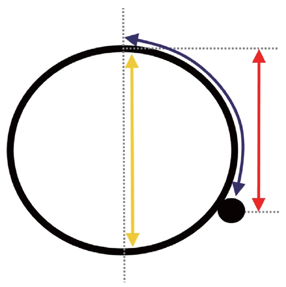
The patients were categorized in two groups as male (group 1) and female (group 2). The operations and measurements of all patients were done by the same surgeon (FA). All the nerves were recognized at the lower pole of thyroid and followed until the level of laryngeal entrance. After the thyroid tissue was completely removed, four measurements were done: (1) trachea vertical height (tvh, vertical length between the anterior and posterior ends of trachea at the level of first tracheal ring tvh) (Fig. 1). (2) Right rln height and left rln height (rrh and lrh, vertical length between the recurrent laryngeal nerves and the line tangent to the tracheal anterior surface at the first tracheal ring) (Fig. 1). (3) Right and left rln to trachea anterior face median raphe distance (rrd and lrd, the distance of right and left recurrent laryngeal nerves from the median raphe at first tracheal ring level on the anterior side of trachea) (Fig. 1). (4) Right and left rln respect to trachea ratio (rrtr and lrtr, the location of the recurrent nerve with respect to first tracheal ring was calculated from the the tracheal vertical length and rln length and formulized). It was named “formula 1” and for example, the formula for right side was: (tvh-rrh)÷tvh. This ratio demonstrated the depth of recurrent nerve at the first tracheal level.
Statistical analysis
The parameters were analyzed statistically with independent t-test using IBM SPSS ver. 19.0 (IBM Co., Armonk, NY, USA). Also, the parameters related to right and left recurrent laryngeal nerves were analyzed with paired sample t-test without differentiating sex. P-value<0.05 was considered statistically significant. Correlations were examined by using the Pearson method and the results were expressed as r-values.
RESULTS
The average age of males and females were 61.50±8.96 and 40.79±15.02 years respectively; this average was significantly higher in males than in females (P<0.002).
Tvh was 2.20±0.28 cm in males and 2.13±0.28 cm in females. The difference was not significant between the two groups (P=0.599). Rrh average was 1.61±0.31 cm in males and 1.50±0.26 cm in females. Rrh scores were not significantly different between two groups (P=0.364). Lrh average was 1.88±0.26 cm in males and 1.63±0.27 cm in females. Lrh scores were significantly higher in males than in females (P=0.040). Rrd average was 2.18±0.67 cm in males and 1.92±0.52 cm in females. The difference was not significant between two groups (P=0.152) (Table 1). Lrd average was 2.38±0.65 cm in males and 2.06±0.33 cm in females. This difference was not significant (P=0.064). Rrtr average was 0.26±0.07 in males and 0.29±0.10 in females and the difference was not significant (P=0.477). Lrtr average was 0.14±0.03 in males and 0.23±0.10 in females. This difference was not significant (P=0.057) (Table 1).
In total patient group; recurrent vertical heights, distance of recurrent laryngeal nerve, trachea level where recurrent laryngeal nerve located on the right and left recurrent laryngeal nerves were compared. The left recurrent laryngeal nerves were significantly deeper than right recurrent laryngeal nerves (lrh, 1.66±0.28 and rrh, 1.52±0.27; P<0.001; Pearson r=0.713). There was a strong positive correlation between lrd (2.10±0.39) and rrd (1.95±0.41) (P<0.001; Pearson r=0.763). Lrd was significantly higher than rrd (P<0.001). There was a strong positive correlation between lrtr (0.22±0.10) and rrtr (0.28±0.09) (P<0.001; Pearson r=0.569). Left recurrent laryngeal nerve was located in a significantly lower segment of the trachea than right recurrent nerve (P<0.001) (Table 2).
DISCUSSION
Thyroid surgery has made great progress since Kocher. However, recurrent laryngeal nerve paralysis is still one of the most significant complications of thyroid surgery despite all advances. In order to avoid this complication, many surgical techniques and landmarks were determined. These studies all aim to decrease the rate of recurrent laryngeal nerve paralysis.
Recurrent laryngeal nerve anatomy is needed to be wellknown and studied. Recurrent laryngeal nerve reaches the level of thyroid lower pole after leaving the vagus and crossing the subclavian artery on right and arcus aorta on left. This level and the course of recurrent laryngeal nerve after this point is very critical in nerve injury during thyroid surgery.
Recurrent laryngeal nerve, at the level of thyroid lower pole, reaches close to trachea on both sides, being more oblique on the right and more medial on the left [4-7]. There are various information about the course of the recurrent laryngeal nerve in relation to trachea after they cross the inferior thyroid artery at the thyroid lower pole level [10]. They most frequently pass through tracheoesophageal groove (59% to 65% on right, 70% to 77% on left), less frequently lateral to the trachea (28% to 33% on right, 17% to 22% on left), and the least frequently anterolateral to the trachea (4% to 8% on right, 3% to 6% on left) [4-7,11].
Several different sources report that recurrent nerves may cross the inferior thyroid artery in the front, back or in between [8,9]. Because of all these variations, inferior thyroid artery is thought to be a reliable anatomic point for positioning recurrent laryngeal nerve. Inferior parathyroid gland is also used as an important landmark at this region [12]. Usually, recurrent laryngeal nerve is located deep under the parathyroid gland. After this point, the recurrent laryngeal nerves direct towards larynx and Berry ligament and branch diversely around the Berry level [13]. The posterior branch here is usually the sensory branch and it merges with the internal branch of the superior laryngeal branch and forms Galen anastomosis [14]. Anterior branch is the one that carries the motor fibers [13,15-18]. This ligament is a critical border point in recurrent laryngeal nerve isolation [7,8]. The nerve can be established at this point even if the nerve is non-recurrent [10]. Besides, this area, especially in case of a growth in thyroid, cancer or thyroiditis, is the most susceptible area for injury [1,8,19].
Again, recurrent nerve damage during total thyroidectomy is mostly seen around Berry ligament [20,21]. The nerve that passes behind and through the ligament enters the larynx around the level of cricoid cartilage behind the muscle [7]. However, the dissection of this area is tricky because of the dense fibrous mesh and vascularity [22]. Furthermore, in this region, the thyroid tissue is very close to the surface of laryngeal cartilage causing difficulty in recurrent laryngeal nerve dissection and causing hemorrhage in thyroid which may end up in recurrent laryngeal nerve paralysis [12]. Also, the extension of thyroid lateral lobes to the posterior of the trachea (Zuckerkandl’s tuberculum) in this area, is one of the factors that raise difficulty to determine the recurrent laryngeal nerve [7,23].
In order to determine the location of recurrent laryngeal nerve in the area under Berry ligament where the nerve is most susceptible to injury, we defined several different anatomic relationships between trachea and the entry point of the recurrent nerve to larynx. For this purpose, we measured the vertical height of the first tracheal ring, vertical height of the recurrent laryngeal nerves at first tracheal ring level on both right and left sides and measured the distance of recurrent laryngeal nerve to the median raphe on the front surface of first tracheal ring on both sides. Additionally, we formulized the level that corresponds to the position of the recurrent laryngeal nerve with respect to the vertical height of the first tracheal ring.
In the study by Liu et al. [24], the trachea was used to locate the recurrent laryngeal nerve and similar to our study, the vertical heights of the recurrent laryngeal nerves were measured. However, different from our study, these measurements were done at the level of third tracheal ring.
The study by Sasou et al. [25] measured the distance between recurrent laryngeal nerve and Berry ligament at trachea midline. This study is especially important as it focuses on the first and second tracheal rings on which the Berry ligament attaches. Furthermore, Berlin [26] and Jatzko et al. [27] studied the anatomic relationship between the Berry ligament and the recurrent laryngeal nerve.
Also, we thought that the first tracheal ring is one of the best ways to evaluate rln position because it is very close to area that the nerve gets into larynx. The nerve has very stable position than the lower segments of trachea like third ring and thight bonds to other structures like Berry ligament.
In our study, although tvh (P=0.599), rrh (P=0.364), and lrh (P=0.040) parameters were higher in males than in females, only the difference in lrh was statistically significant. This showed that the recurrent nerves on left sides of males patients were deeper than the female patients. When rrd (P=0.152), lrd (P=0.064), rrtr (P=0.477), and lrtr (P=0.057) parameters were compared in both groups, rrd and lrd parameters were higher in males, whereas rrtr and lrtr parameters were lower in males. However, these differences were not statistically significant. In the study by Liu et al. [24] which recurrent laryngeal nerve vertical height was examined, at the level of third tracheal ring, the recurrent laryngeal nerve height was higher in males both in cadavers and clinical cases. In our study, although the measurements were done in the first tracheal ring level, the data was deemed consistent.
There are several important points arised from the results of our study. In all cases, the midpoint of tvh at the first tracheal ring emerged to be an important landmark from the calculations of trachea-recurrent ratio (lrtrr and rrtr). Recurrent laryngeal nerve on both right and left sides were found to be inferior to the midpoint of tvh (we named this point as “Akil point”) and this showed that the upper half of tvh was safe during surgery. The lrtr analysis showed that left recurrent nerves were significantly deeper at the first treacheal ring and located lower at the posterior half of the first tracheal ring (P<0.001). Secondly, in all male patients and 12 of female patients, right recurrent laryngeal nerve was found significantly more superficial than the left recurrent laryngeal nerve at the first tracheal ring level. When right and left sides were compared without sex discrimination, lrh was located significantly deeper than rrh (P<0.001). Similarly, in all male patients and 12 of female patients, the distance between recurrent laryngeal nerve and the trachea anterior face median raphe at the first tracheal ring level is found to be less on the right side than the left side. When the midline distances were compared without sex discrimination, the left recurrent laryngeal nerve was significantly distant than the right to trachea midline (P<0.001).
Although the small sample size of the patient pool is a disadvantage, we believe that the landmarks determined in this study and the ease it provides in recurrent laryngeal nerve identification in thyroid surgery can help to decrease the recurrent laryngeal nerve injury rates.
In conclusion, this study reports that the recurrent laryngeal nerve is always located at the lower half of the trachea vertical height at the first tracheal ring level and the left recurrent laryngeal nerve is located significantly deeper than the right in all males and in most females. The study also formulates the distance of the recurrent laryngeal nerves to the anterior face median raphe of the trachea. We believe this study is adds valuable data on the anatomy of the area where most damage occurs in the recurrent laryngeal nerve. We expect the results of this study to be examined thoroughly by further studies and these measurements will help to decrease the rates of recurrent laryngeal nerve paralysis.
HIGHLIGHTS
▪ Recurrent laryngeal nerve on both sides are located inferiorly to the midpoint of trachea vertical height at first tracheal cartilage level.
▪ Upper half of trachea vertical height at first tracheal cartilage level is safe during surgery.
▪ Left recurrent laryngeal nerves are significantly deeper at the first treacheal ring.
▪ Left recurrent laryngeal nerves located lower at the posterior half of the first tracheal ring.