Factors Affecting the Extrusion Rate and Complications After Ventilation Tube Insertion: A Multicenter Registry Study on the Effectiveness of Ventilation Tube Insertion in Pediatric Patients With Chronic Otitis Media With Effusion—Part II
Article information

Abstract
Objectives
The impacts of ventilation tube (VT) type and effusion composition on the VT extrusion rate and complications in children with otitis media remain unclear. This part II study evaluated the factors affecting the extrusion rate, recurrence rate, and complications of VT insertion.
Methods
A prospective study was conducted between June 2014 and December 2016 (the EVENT study [analysis of the effectiveness of ventilation tube insertion in pediatric patients with chronic otitis media]), with follow-up data collected until the end of 2017. Patients aged <15 years diagnosed with otitis media with effusion who received VT insertion were recruited at 15 tertiary hospitals. The primary outcomes were time to extrusion of VT, time to effusion recurrence, and complications.
Results
Data from 401 patients were analyzed. After excluding the results of long-lasting tubes (Paparella type II and T-tubes), silicone tubes (Paparella type I) exhibited a significantly longer extended time to extrusion (mean, 400 days) than titanium tubes (collar-button-type 1.0 mm: mean, 312 days; P<0.001). VT material (hazard ratio [HR], 2.117, 95% confidence interval [CI], 1.254–3.572; P=0.005), age (HR, 3.949; 95% CI, 1.239–12.590; P=0.02), and effusion composition (P=0.005) were significantly associated with the time to recurrence of middle ear effusion. Ears with purulent (mean, 567 days) and glue-like (mean, 588 days) effusions exhibited a shorter time to recurrence than ears with serous (mean, 846 days) or mucoid (mean, 925 days) effusions. The revision VT rates during follow-up were 3.5%, 15.5%, 10.4%, and 38.9% in ears with serous, mucoid, glue-like, and purulent effusions, respectively (P<0.001). The revision surgery rates were higher among patients aged <7 years than among those aged ≥7 years.
Conclusion
Silicone tubes (Paparella type I) were less prone to early extrusion than titanium 1.0 mm tubes. VT type, patient age, and effusion composition affected the time to recurrence of effusion.
INTRODUCTION
Otitis media is the most common cause of acquired hearing loss in the pediatric population, affecting two-thirds of all children within the first 3 years of life [1]. As most cases of otitis media resolve spontaneously, watchful waiting or antibiotics may be sufficient to alleviate this disease. However, surgical interventions (myringotomy and ventilation tube [VT] insertion) are required for prolonged chronic otitis media with effusion (OME) or recurrent acute otitis media (AOM) [2]. VT insertion under general anesthesia is the most commonly performed surgery in the pediatric population [3]. In most cases, inserted tubes extrude naturally after 6–18 months, except for long-lasting tubes (such as Paparella type II and Goode T-tubes) that persist for ≥2 years [4]. Factors including status of middle ear effusion, history of previous VT surgery, tympanic membrane status, repeated episodes of otorrhea, location of myringotomy incision, and type of VT affect the time to VT extrusion [5-7]. Song et al. [6] reported that revision VT insertion and serous effusion were associated with a shorter time to extrusion, but age and tympanic membrane status did not significantly affect the time to VT extrusion. In contrast, Leopold and McCabe [8] reported that general anesthesia, initial surgery, and younger age (<9 years old) were associated with longer VT retention. A recent multicenter study demonstrated that tympanic membrane status was correlated significantly with the tube retention duration and recurrence rate [5], highlighting the discrepancies in the literature.
Various types of VTs with different sizes, shapes, coatings, and materials are currently available [9]. The time to tube extrusion depends on the inner flange diameter and shape. Short-lasting tubes are typically inserted for initial cases, whereas long-lasting tubes are selected for patients with a history of multiple VT insertions or children with systemic difficulties with repeated general anesthesia. Titanium tubes are increasingly being used due to their high biocompatibility [10,11]. Nevertheless, there is a paucity of literature on the influence of tube shape, design, and material on extrusion time and time to recurrence.
The complications associated with VT insertion include post-tube otorrhea, tube plugging (obstruction), granulation tissue formation, cholesteatoma development, and persistent tympanic membrane perforation [12]. Middle ear effusion composition affects the tube plugging rate, and longer tube retention is associated with higher persistent perforation and infection rates [13-15]. However, a randomized study using four different tube types reported no significant differences in the persistent perforation rate between short- and long-lasting tubes [16]. However, multiple VT insertion history is associated with a higher rate of persistent perforations [14]. These discordant findings highlight the lack of consensus regarding factors associated with complication rates after VT insertion.
To address these gaps, this multicenter registry study (EVENT study [an analysis of the effectiveness of ventilation tube insertion in pediatric patients with chronic otitis media: a multicenter registry study]) aimed to investigate the effectiveness of VT insertion in pediatric patients with chronic otitis media. Part I of the EVENT study previously reported the microbiology of OME in children [17]. This study (part II of the EVENT study) describes the factors affecting times to VT extrusion and effusion recurrence, as well as post-VT insertion complications.
MATERIALS AND METHODS
Study design
From June 2014 to December 2016, patients with OME who received VT insertion in 15 tertiary hospitals throughout Korea were prospectively enrolled, as previously reported [17]. Followup data of the enrolled patients were collected until December 2017. Inclusion criteria were as follows: (1) age <15 years and presented with OME requiring VT insertion; (2) OME diagnosis based on the presence of middle ear effusion without signs of acute inflammation; and (3) diagnosis performed with an otoscope, pneumatic otoscope, microscope, endoscope, and/or tympanometer type B/C. Patients were excluded if they or their guardians declined enrolment. Bacterial cultures of middle ear effusions were recommended during VT insertion. Data from the hospital records of patients who agreed to participate in the study were collected [17].
The study protocol was reviewed and approved by the Institutional Review Boards of all participating hospitals. This protocol was registered with the Clinical Research Information Service (http://cris.nih.go.kr, No. KCT0001378) for public access. Written informed consent was obtained from all parents/guardians and children aged >7 years. Data were registered at each hospital using Internet-based electronic case report forms (eCRFs; http://otology.crf.kr).
Statistical methods
The primary outcomes were times to VT extrusion and effusion recurrence as well as postoperative complications (revision VT insertion during follow-up and tube otorrhea, persistent perforation, and tube plugging rates). The “time to VT extrusion” was calculated as the midpoint between the last date when VT was found to be in place and the first date when VT was found to be extruded. The “time to effusion recurrence” was calculated as “the period after the surgery until the recurrence of effusion.” Audiometric data were used as secondary outcomes. Statistical analyses were performed using IBM SPSS ver. 22.0 (IBM Corp., Armonk, NY, USA). Categorical variables were analyzed with Pearson’s chi-square or Fisher’s exact tests. Descriptive data were shown as means and standard deviations. Times to VT extrusion and effusion recurrence according to different factors were presented using Kaplan-Meier survival plots (estimated mean and 95% confidence intervals [CI]) and were compared using the log-rank test. Contributory factors in surgical success were assessed using multivariate Cox regression analyses; a P-value of 0.1 was the inclusion criterion for multivariate analysis. Patients with a follow-up duration of <6 months were excluded from the analysis of factors associated with complications after VT insertion. Statistical significance was set at P<0.05.
RESULTS
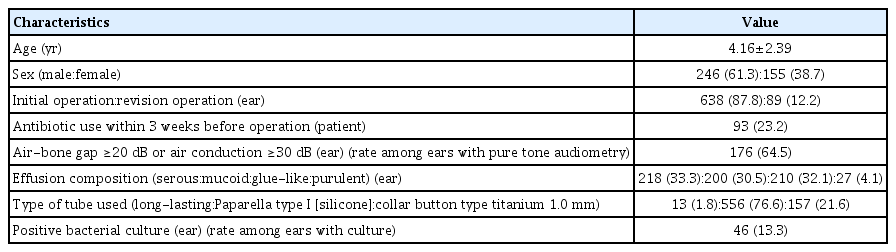
Among the 432 enrolled patients, data from 401 patients (727 operated ears) were analyzed after excluding patients with missing data. Of these, 246 (61.3%) were male, with a mean age of 4.16±2.39 years. The mean follow-up period after VT insertion was 331±238 days (range, 3–1,377 days). The first follow-up was mostly conducted within 3 weeks of surgery, and the second follow-up within 6 months after VT insertion. In total, 22% (89 cases) were revisions with a history of VT insertion (Table 1). In the analysis of complication rates, 489 ears remained after excluding patients with <6 months of follow-up.
Patient characteristics
Long-lasting tubes (Paparella type II and Goode T-tubes; both composed of silicone) were inserted in 13 ears. The time to extrusion after tube insertion was significantly longer for long-lasting tubes (mean, 678 days; 95% CI, 476–880 days) than for other tubes (Paparella type I silicone and titanium collar-button-type tubes; mean, 378 days; 95% CI, 359–396 days; P=0.001). Ears with long-lasting tubes were excluded from further analysis because the persistent perforation rate after extrusion was significantly higher for long-lasting tubes than for short-lasting tubes (20% vs. 0.8%; P=0.005) (Supplementary Table 1).
Univariate analysis revealed that time to extrusion was significantly longer for silicone tubes (Paparella type I, inner diameter: 1.14 mm, Medtronic Xomed, Jacksonville, FL, USA; inserted in 556 ears) than for titanium tubes (inner diameter: 1.0 mm, collar-button-type, Spiggle & Theis, Overath, Germany; inserted in 157 ears) (mean, 400 vs. 312 days; P<0.001) (Fig. 1A). Patients <7 years old exhibited a significantly shorter time to VT extrusion compared to patients aged ≥7 years old (P=0.043). No significant differences in the time to effusion recurrence were noted for other factors in the univariate analysis, including the side of insertion, sex, preoperative antibiotic use, revision case, effusion status, poor hearing (defined as air-bone gap ≥20 dB or air conduction ≥30 dB), and positive bacterial growth in effusion cultures (Table 2).
Cox hazard ratio model. (A) Time to tube extrusion according to the type of material (in short-lasting tubes). Silicone (Paparella type I) tubes exhibited a significantly longer time to extrusion than titanium tubes (1.0 mm collar-button-type). (B) Time to effusion recurrence according to age. The time to effusion recurrence was significantly different between patients aged <7 years and those aged ≥7 years. (C) Time to effusion recurrence according to the type of material. Silicone tubes exhibited a significantly longer time than titanium tubes. (D) Time to effusion recurrence according to middle ear effusion composition. Ears with serous (846 days) and mucoid (925 days) effusions exhibited significantly delayed recurrence compared to those with glue-like (588 days) or purulent (567 days) effusions.

Univariate analysis of factors affecting times to extrusion and effusion recurrence
Univariate analysis revealed that patients <7 years old exhibited significantly more frequent early recurrence (P=0.002) (Fig. 1B). The time to recurrence was significantly longer for silicone tubes than for titanium tubes (mean, 985 vs. 557 days; P<0.001) (Fig. 1C). Effusion status exhibited significant differences in time to effusion recurrence (mean for serous effusion: 846, mucoid: 925, glue-like: 588, and purulent: 567 days) (Fig. 1D). No significant differences in time to tube extrusion were observed for other factors, including the side of insertion, sex, preoperative antibiotic use, revision case, poor hearing, and positive bacterial culture (Table 2).
Multivariate analysis using a Cox proportional hazards model was conducted to evaluate the factors influencing the times to extrusion and effusion recurrence. The VT material was the only independent predictor of time to VT extrusion (hazard ratio [HR], 1.746; P<0.001) (Table 3). VT material (HR, 2.117; P=0.005), age >7 years (HR, 3.949; P=0.02), and effusion composition (P=0.005) were independent predictors of the time to effusion recurrence. The time to OME recurrence was significantly shorter for glue-like and purulent effusions than for serous and mucoid effusions (Table 4).

Multivariate analysis of factors affecting time to VT extrusion (n=713 ears)

Multivariate analysis of factors affecting time to effusion recurrence (n=713 ears)
The incidence of tube otorrhea was 2.9% and 0.9% in the silicone tube and titanium tube groups, respectively, with no significant between-group difference (Supplementary Table 2). Tube plugging by crust or bloody clots was observed in 29 ears (4.0%) during follow-up. The plugging rate was 7.8% for titanium tubes and 3.8% for silicone tubes; this difference was not statistically significant (P=0.075). Persistent perforation after extrusion was observed in four (1.1% of 373 ears) silicone tube-inserted ears, but not in titanium tube-inserted ears; this difference was not statistically significant (P>0.05, Fisher’s exact test) (Supplementary Table 2). The rates of revision surgery during follow-up were significantly higher in patients aged <7 years than in those aged ≥7 years (Supplementary Table 3). The rate of persistent perforation after extrusion was 2.2% in revision VT insertion ears and 0.5% in initial VT insertion ears, which did not reflect a statistically significant difference (P=0.156, Fisher’s exact test) (Supplementary Table 4). The rates of revision VT insertion surgery were higher for ears with an initial tube than for ears with a previous history of VT insertion during follow-up (20.2% vs. 9.6%, P=0.005).
No significant differences were noted in the rates of tube otorrhea and tube plugging between initial and revision cases (Table 4). The revision VT insertion rates during follow-up were 3.5%, 15.5%, 10.4%, and 38.9% for serous, mucoid, glue-like, and purulent effusions, respectively, which was a statistically significant difference (P<0.001) (Table 5). The postoperative air-conduction thresholds were significantly lower than the preoperative air-conduction thresholds (mean, 19.28±16.52 dB vs. 28.53±14.01 dB; P<0.001). Preoperative antibiotic use (Supplementary Table 5), intraoperative culture results (Supplementary Table 6), and preoperative hearing (Supplementary Table 7) did not significantly affect the complication rate after VT insertion. No cholesteatoma or granulation formation was observed in any patients.
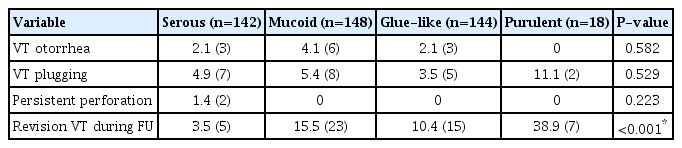
Complication rates according to middle ear effusion composition
DISCUSSION
Myringotomy with VT insertion is the most common surgery performed in children, accounting for up to 600,000 cases annually in the United States [12]. Clinical practice guidelines recommend bilateral VT insertion for bilateral OME lasting for >3 months and for cases with documented hearing loss or frequently recurring AOM (three episodes in 6 months or four episodes in 1 year) [2,7]. Recent meta-analyses and systematic reviews have suggested that VTs improved hearing during the early period (1-3 months) of OME compared to watchful waiting, but there was no evidence of benefit at 12–24 months [7,18]. Djordjevic et al. [19] reported that 51% of patients experienced at least one long-term complication of VT insertion (evaluated at 22–27 years after the initial VT insertion), including atrophic tympanic membrane scarring, tympanosclerosis, tympanic membrane retraction, persistent perforations, and sensorineural hearing loss. Accordingly, VT insertion should be performed using the appropriate type of tube after weighing the potential benefits and risks in consideration of each patient’s condition.
VTs typically contain an inner flange to avoid extrusion and an outer flange to avoid intrusion into the middle ear. The inner flange diameter and shape affect the time to extrusion [20]. With the exception of long-lasting tubes, which have large or T-shaped inner flanges (such as Paparella type II or Goode T-tubes, respectively), VTs extrude naturally in most cases. If extrusion occurs too early (earlier than 12 months), OME may recur following natural healing of the tympanic membrane, potentially requiring revision surgery. In contrast, permanent tympanic membrane perforation rates increase after 18 months of tube maintenance [13]. Hence, the ideal tube for initial surgery should remain dry without complications (tube plugging or otorrhea) for >12 months and should spontaneously extrude before 18 months without persistent perforation.
Insufficient evidence currently exists regarding the effects of tube design and material on extrusion. Gibb and Mackenzie [21] reported that tube design was the only significant factor affecting VT extrusion. One study reported that titanium tubes were extruded more slowly than Shepard Teflon tubes [10], and C-Flex tubes were extruded more slowly than silicone tubes. However, no study to date has directly compared titanium and silicone tubes [4]. In our study, Paparella type I tubes exhibited a significantly longer time to extrusion than titanium collar-button-type tubes. As VTs with larger inner flanges extrude later, this difference may be due to the larger inner flange of silicone Paparella type I tubes. However, Paparella type I tubes contain a notch in the inner flange, which facilitates tube insertion through the myringotomy site. This likely influences extrusion time by reducing resistance to outward forces of the tympanic membrane.
Material hardness may also affect tube extrusion time. Kim et al. [4] proposed that the soft texture of silicone, which has poor mechanical strength, contributes to its earlier extrusion than the firmer C-Flex tubes. However, we observed that titanium tubes, which are harder than other materials, were extruded even earlier than silicone tubes. This may have been due to different inner flange shapes and diameters, apart from material hardness and other characteristics. Therefore, our data are insufficient to conclude that titanium VTs are extruded earlier than silicone VTs, and further studies using VTs of the same shape with different materials will be needed.
In this study, among patients who received short-lasting tubes, the mean time to VT extrusion was 378 days (95% CI, 359–396 days), and the mean time to recurrence of effusion was 892 days (95% CI, 787–997 days). Between that period (mean duration between VT extrusion and recurrence of effusion: 514 days), 90 ears (12.6%) showed extrusion of the VT. Therefore, patients may be recommended to receive follow-up for at least 1–2 years after VT extrusion to monitor the recurrence of OME. Furthermore, Paparella type I silicone tubes displayed significantly delayed effusion recurrence compared to titanium collar-button-type tubes in our study. Indeed, the early recurrence of OME after VT surgery is a significant prognostic factor for multiple VT insertions [22]. Hence, Paparella type I tubes may be preferred over titanium tubes for OME due to their favorable indwelling duration.
Song et al. [6] reported that effusion composition during surgery was an independent predictor of tube extrusion time. In contrast, we did not observe a significant difference in tube extrusion time according to effusion composition, consistent with the results of Han et al. [23]. A recent study also reported that viscosity of effusions, as measured by a viscometer, did not influence the extrusion time of VT [24]. Therefore, the tube extrusion time seems to be independent of the effusion composition or viscosity in OME.
Conflicting results on the relationship between effusion composition and multiple tube insertions have been reported. Choi et al. [22] reported that serous effusions increased the risk of multiple VT insertions. However, Salam and Wengraf [25] found that mucoid effusions were associated with a higher recurrence rate. Other studies reported no significant relationship between effusion composition and OME recurrence [26,27]. We observed a significant difference in the time to effusion recurrence after tube extrusion according to effusion composition. Ears with serous and mucoid effusions exhibited significantly delayed recurrence compared to those with glue-like or purulent effusions. This trend implies that glue-like or purulent effusions suggest a more advanced stage of OME than serous and mucoid effusions and lead to an earlier recurrence of OME. Future studies should evaluate the effects of middle ear effusion on OME recurrence.
The second or multiple VT placement rates in children are as high as 20%–50% [22]. In previous reports, revisions exhibited a significantly shorter extrusion time [6,8,22]. However, in our study, revisions (mean, 345 days; 95% CI, 305–385 days) did not show a significantly shorter extrusion time than initial cases (mean, 387 days; 95% CI, 366–408 days; P=0.097). Moreover, no significant differences were observed in the time to effusion recurrence between ears with initial and revision VT insertion. Therefore, surgeons can expect the usual course of VT even in revision cases.
Univariate analysis revealed that the times to extrusion and effusion recurrence were significantly shorter in patients aged <7 years old than in their older counterparts. However, in multivariate analysis, only the time to effusion recurrence was significantly different between patients aged <7 years old and those aged ≥7 years. This corresponds with the findings of previous studies that reported no significant difference in the time to extrusion according to age [6,21]. A shorter time to effusion recurrence before the age of 7 years may be a natural occurrence, given the decrease in OME incidence after the age of 7–8 years [28].
Obstruction of the inner lumen of VTs by crust or blood clots (7%–10% of cases) is frequently observed during post-surgical follow-up [15,29]. Plugging may perturb middle ear ventilation, resulting in hearing loss or even early VT extrusion [4]. The narrower inner lumen of titanium tubes (1 mm vs. 1.14 mm) may lead to a higher incidence of tube obstruction. However, our results demonstrated that the VT obstruction rate was not significantly different between titanium and silicone tubes. Conrad et al. [29] reported that serous effusions exhibited the highest rate of tube plugging. In contrast, our data showed that purulent effusions demonstrated the highest rate of tube plugging, with no significant differences between effusion compositions. Our findings imply that VT plugging is not influenced by the effusion composition during the operation.
Late tube otorrhea after VT insertion is also common, with 0.36–3.6 episodes per child each year [20]. A randomized controlled study by Knutsson et al. [16] reported that the duration to first infection was significantly longer for silicone than for fluoroplastic tubes, which was attributed to the high biocompatibility of silicone. Titanium is also biocompatible and is a material for various in-body implants, including ossiculoplasty replacement prostheses. In our study, the incidence of VT otorrhea was not significantly different between titanium tubes and silicone tubes. Bacterial biofilm formation on VTs has been proposed as a cause of intractable tube otorrhea, particularly at the round rims of Paparella-type tubes. Studies have assessed the antibiofilm coatings of tubes, but their clinical efficacy remains a matter of debate [30].
We observed a significantly higher rate of persistent tympanic membrane perforations for long-lasting tubes than for short-acting tubes, consistent with previous reports [14,18,20]. Titanium tubes did not exhibit persistent perforations after extrusion, whereas silicone tubes exhibited a 1.1% perforation rate. Longer tube maintenance results in a higher persistent perforation rate, and the longer retention of silicone tubes than that of titanium tubes may have resulted in a higher frequency of perforation [15]. In long-term follow-up, the rate of tympanic membrane abnormalities was significantly higher in patients with multiple VT insertions than in patients with a single VT insertion [1,14]. However, we did not observe significantly higher rates of persistent perforation in revisions than in initial cases. Further research is required to evaluate the association of multiple tube insertion with persistent perforation.
The strength of this study is that we enrolled a large population from multiple centers and prospectively collected data using eCRFs. The main limitation of our study is that the specific tube type was selected according to each surgeon’s preference. Differences in surgical techniques (e.g., incision and tube placement location) and follow-up cycles among surgeons may have influenced the postoperative results. Further prospective randomized controlled trials are warranted to determine the factors affecting the VT extrusion time and time to OME recurrence.
Compared to titanium 1.0 mm collar-button tubes, silicone Paparella type I VTs were less prone to early extrusion. The type of VT, the patient’s age, and effusion composition affected the time to effusion recurrence. Therefore, the appropriate VT should be selected considering the patient’s condition.
HIGHLIGHTS
▪ In this multicenter registry study of 401 pediatric patients, the time to extrusion was longer for silicone tubes than for titanium tubes.
▪Ventilation tube (VT) type, patient age, and effusion composition affected the time to recurrence of effusion after VT.
▪The rates of revision surgery during follow-up were significantly higher in patients aged <7 years than in those aged ≥7 years.
▪An appropriate type of VT should be selected for otitis media with effusion according to the patient’s condition.
Notes
No potential conflict of interest relevant to this article was reported.
AUTHOR CONTRIBUTIONS
Conceptualization: JC, JWC, HJP, KYL. Data curation: MHY, YSC, JC, JHC, GCH, BCJ, ISM, SHL, JR, SGY. Formal analysis: MHY, JC, JWC, KYL, JHS. Funding acquisition: YSC, YHC, JWC, JHL, SGY. Methodology: MHY, JHC, GCH, BCJ, DKK. Project administration: MHY, JC, JWC, GCH, SHL. Visualization: MHY, BCJ, DKK, KSK, JHL, ISM, HJP, SNP, JHS, SGY. Writing–original draft: MHY, JWC. Writing–review & editing: YSC, JC, YHC, JHC, JWC, GCH, BCJ, DKK, KSK, JHL, KYL, SHL, ISM, HJP, SNP, JR, JHS, SGY.
Acknowledgements
This work was supported by the Otitis Media Research Project funded by the Korean Otologic Society, Seoul, Republic of Korea and a National Research Foundation of Korea (NRF) grant funded by the Korean government (Ministry of Science and ICT) (No. 2022R1C1C1010262).
SUPPLEMENTARY MATERIALS
Supplementary materials can be found online at https://doi.org/10.21053/ceo.2022.00934.
Complication rates according to long-lasting vs. short-lasting tubes
Complication rates according to type of ventilation tube
Complication rates according to age
Complication rates according to initial vs. revision operation
Complication rates according to preoperative antibiotic use
Complication rates according to intraoperative culture results
Complication rates according to preoperative hearing